Bacillus sp. A8a reduces leaf wilting by Phytophthora and modifies tannin accumulation in avocado
By
Edgar Guevara Avendaño,
Itzel Anayansi Solís García,
Alfonso Méndez Bravo,
Fernando Pineda García,
Guillermo Angeles Alvarez,
Carolina Madero Vega,
Sylvia P. Fernández Pavía,
Alejandra Mondragón Flores,
Frédérique Reverchon *
* Corresponding Author. Email: - / Institution: Instituto de Ecología, A.C.
Received: 17/September/2023 – Published: 08/December/2023 – DOI: https://doi.org/10.18781/R.MEX.FIT.2309-2
Abstract Background/Objective. The objective was to assess the biocontrol capacity of Bacillus sp. A8a in avocado (Persea americana) plants infected by Phytophthora cinnamomi.
Materials and Methods. A greenhouse experiment was implemented with four treatments: 1) control plants; 2) plants infected with P. cinnamomi; 3) plants inoculated with Bacillus sp. A8a; 4) plants infected with P. cinnamomi and inoculated with Bacillus sp. A8a. We evaluated several morpho physiological variables during the experiment, which lasted 25 days after infection (dai). Moreover, we analyzed tannin density in stems at 25 dai to determine the plant defense response against the disease.
Results. Inoculation with strain A8a reduced wilting symptoms by 49 % at 25 dai, compared with non-inoculated plants. No differences were detected in morpho physiological variables between treatments. However, a greater tannin accumulation was registered in the xylem of infected plants, whilst plants inoculated with strain A8a displayed a larger tannin density in the cortex.
Conclusion. Our results confirm the biocontrol activity of Bacillus sp. A8a in avocado plants and suggest that tannin differential accumulation in the cortex of plants inoculated with the bacteria may contribute to the enhanced tolerance of avocado plants against Phytophthora root rot.
Keywords:
Biocontrol, Chemical defense, Persea americana, Phytophthora cinnamomi, Water potential

Figure 1. Effect of the inoculation of Bacillus sp. A8a and P. cinnamomi in avocado trees at 1, 4, 7, 13 and 25 dai (days after infection). A) Percentage of wilting caused by P. cinnamomi; B) tree height (cm); C) stem diameter (cm). Bars show the average of the data (n = 6, treatments C and Pc; n = 3, treatments B and BPc) ± s.d. Different letters indicate significant differences (two-way ANOVA, P ≤ 0.05). In Figure 1A, (+) indicates significant differences between times, compared with (-), for the Pc treatment (ANOVA, P ≤ 0.05). D) Representative photographs of the aerial parts and root systems of avocado trees at 25 dai, in the four treatments. C: control; Pc: infected with P. cinnamomi; B: inoculated with Bacillus sp. A8a; BPc: infected with P. cinnamomi and inoculated with Bacillus sp. A8a.

Figure 2. Leaf water potential in avocado trees at 1, 4, 7, 13 and 25 dai. A) Leaf water potential (MPa) at 5:00 am; B) foliar water potential at 14:00 pm. Bars show the average of the data (n = 3 ± s.d.). Different letters indicate significant differences between treatments at the same time. The water potential measured at 1, 4 and 25 dai was analyzed with a oneway ANOVA and Tukey’s test (P ≤ 0.05). Non-parametric data recorded at 7 y 13 dai were analyzed with KruskalWallis and Wilcoxon rank sum tests ( P ≤ 0.05).

Figure 3. Tannin accumulation in crosssections of the cortex (C), outer xylem and inner xylem of avocado stems from the four treatments at 25 dai. A – C: Control treatment. A. A few tanniniferous cells (T) are observed interspersed in the tissue. B. Tanniniferous cells (T) are observed in the ray parenchyma. Vessels (V) and rays (r) are indicated. C. A few tanniniferous cells (T) are observed in the axial parenchyma and in some radial parenchyma (r). Ph = secondary phloem. Scale: A - C. 30 μm. Figures D – F: treatment Pc. D. Abundant tanniniferous cells (T) are observed. E. Tanniniferous cells (T) are in the radial as well as in the axial parenchyma. V = vessels. F. Tanniniferous cells (T) are mostly concentrated in the radial cells. Scale: D. 60 μm. E and F. 30 μm. Figures G – I: treatment B. G. Cross-section through the secondary phloem (Ph) and cortex (C). Some tanniniferous cells are interspersed amid parenchyma cells full with starch (asterisks). H. A long radial chain of vessels (V) can be observed in the center of the image. Only a few tanniniferous cells (T) are observed in the radial parenchyma. I. Tanniniferous cells (T) are even scarcer than in the outer xylem. Scale: G. 25 μm. H and I. 30 μm. Figures J – L: treatment BPc. J. Tanniniferous cells (T) are observed. K. Grouped or solitary vessels (V) and some tanniniferous cells (T) are observed in the radial parenchyma. L. Few tanniniferous cells (T) are observed in the axial parenchyma. Vessels (V) are grouped in radial chains or in groups of four. Scale: J - L. 35 μm.

Table 1. Photosynthetic rate, stomatal conductance and transpiration in avocado trees infected with P. cinnamomi and inoculated with Bacillus sp. A8a

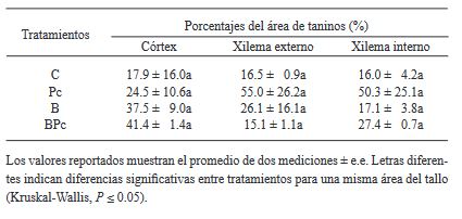
Table 2. Percentage of the area occupied by tanniniferous cells at 25 dai in differ ent stem sections of avocado trees infected by P. cinnamomi and inocu lated with Bacillus sp. A8a.

